Welcome to the Amira-Avizo Software Use Case Gallery
Below you will find a collection of use cases of our 3D data visualization and analysis software. These use cases include scientific publications, articles, papers, posters, presentations or even videos that show how Amira-Avizo Software is used to address various scientific and industrial research topics.
Use the Domain selector to filter by main application area, and use the Search box to enter keywords related to specific topics you are interested in.
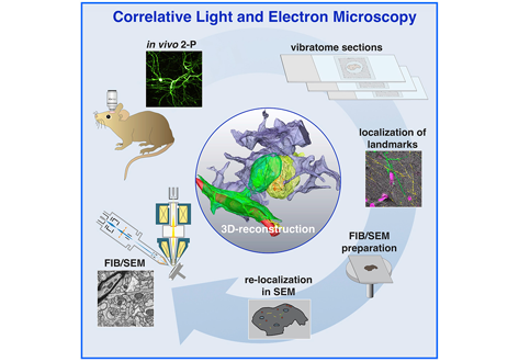
Label-free 3D-CLEM using endogenous tissue landmarks
We demonstrate feasibility of the workflow by combining in vivo 2-photon microscopy and focused ion beam scanning electron microscopy (FIB/SEM) to dissect the role of astrocytic coverage in the persistence of dendritic spines.
Emerging 3D correlative light and electron microscopy (CLEM) approaches enable studying neuronal structure-function relations at unprecedented depth and precision. However, established protocols for the correlation of light and electron micrographs rely ... Read more
Manja Luckner,Steffen Burgold, Severin Filser, Maximilian Scheungrab, Yilmaz Niyaz, Eric Hummel, Gerhard Wanner, Jochen Herms
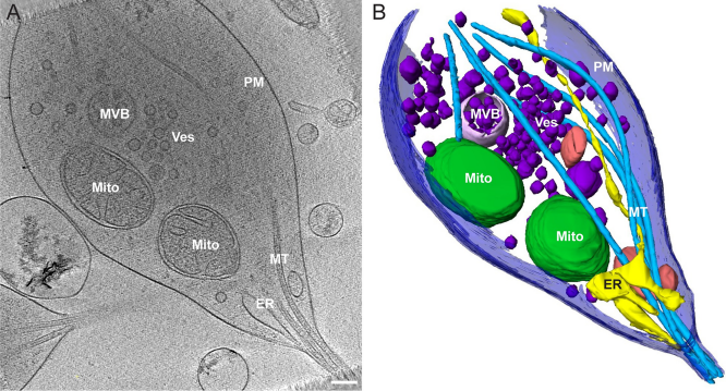
Morphology of mitochondria in spatially restricted axons revealed by cryo-electron tomography
Neurons project axons to local and distal sites and can display heterogeneous morphologies with limited physical dimensions that may influence the structure of large organelles such as mitochondria. Using cryo-electron tomography (cryo-ET), we characterized native environments within axons and presynaptic varicosities to examine whether spatial restrictions within these compartments influence the morphology of mitochondria. Segmented tomographic reconstructions revealed distinctive morphologi... Read more
Tara D. Fischer, Pramod K. Dash, Jun Liu, M. Neal Waxham
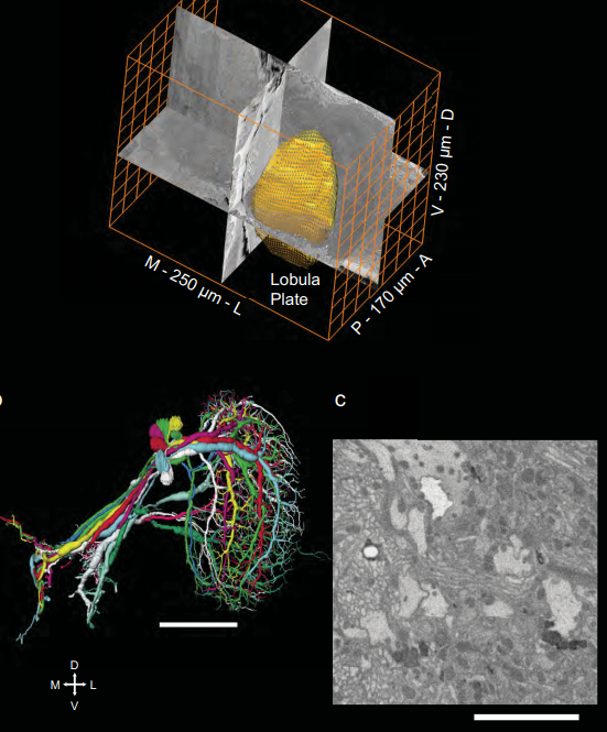
Full reconstruction of large lobula plate tangential cells in Drosophila from a 3D EM dataset
With the advent of neurogenetic methods, the neural basis of behavior is presently being analyzed in more and more detail. This is particularly true for visually driven behavior of Drosophila melanogaster where cell-specific driver lines exist that, depending on the combination with appropriate effector genes, allow for targeted recording, silencing and optogenetic stimulation of individual cell-types. Together with detailed connectomic data of large parts of the fly optic lobe, this has rece... Read more
Kevin M. Boergens , Christoph Kapfer, Moritz Helmstaedter, Winfried Denk, Alexander Borst

Cell-type specific innervation of cortical pyramidal cells at their apical tufts
We investigated the synaptic innervation of apical tufts of cortical pyramidal cells in a region between layers 1 and 2 using 3-D electron microscopy (3D-EM) applied to four cortical regions in mouse. Across all cortices, we found the relative inhibitory input at the apical dendrite’s main bifurcation to be more than 3-fold stronger for layer 2 pyramidal cells than for all other pyramidal cells. Towards the distal tuft dendrites in upper layer 1, however, the relative inhibitory input was a... Read more
Ali Karimi, Jan Odenthal, Florian Drawitsch, Kevin M. Boergens, Moritz Helmstaedter
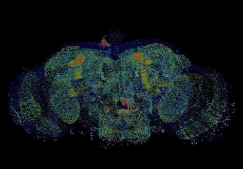
Combined expansion and lattice light sheet microscopy enables high
speed, nanoscale molecular imaging of neural circuits over large volumes.
Optical and electron microscopy have made tremendous inroads in understanding the complexity of the brain, but the former offers insufficient resolution to reveal subcellular details and the latter lacks the throughput and molecular contrast to visualize specific molecular constituents over mm-scale or larger dimensions. We combined expansio... Read more
Ruixuan Gao, Shoh M Asano, Srigokul Upadhyayula, Igor Pisarev, Daniel E Milkie, Tsung-Li Liu, Ved Singh, Austin Graves, Grace H Huynh, Yongxin Zhao, John Bogovic, Jennifer Colonell, Carolyn M Ott, Christopher Zugates, Susan Tappan, Alfredo Rodriguez, Kishore R Mosaliganti, Sean G Megason, Jennifer Lippincott-Schwartz, Adam Hantman, Gerald M Rubin, Tom Kirchhausen, Stephan Saalfeld, Yoshinori Aso, Edward S Boyden, Eric Betzig
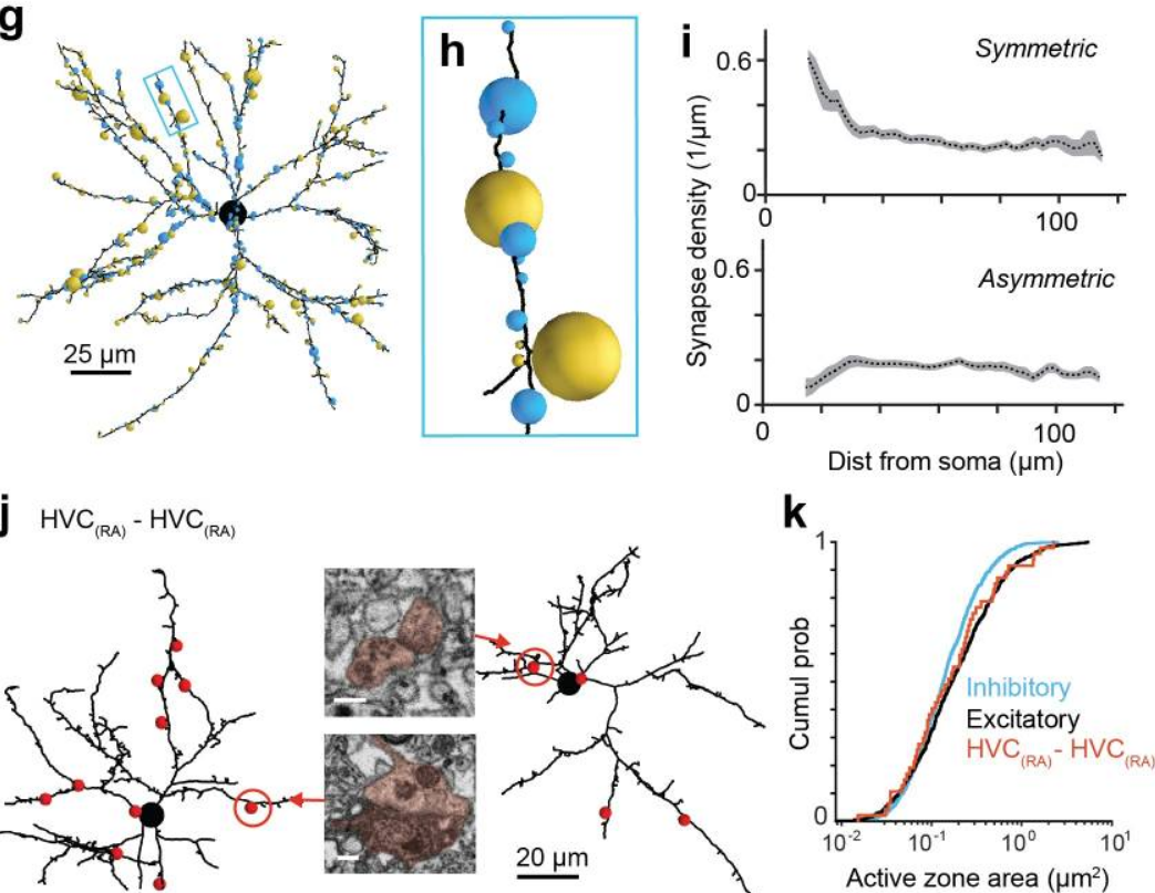
EM connectomics reveals axonal target variation in a sequence-generating network
The sequential activation of neurons has been observed in various areas of the brain, but in no case is the underlying network structure well understood. Here we examined the circuit anatomy of zebra finch HVC, a cortical region that generates sequences underlying the temporal progression of the song. We combined serial block-face electron microscopy with light microscopy to determine the cell types targeted by HVC(RA) neurons, which control song timing. Close to their soma, axons... Read more
Jörgen Kornfeld, Sam E Benezra, Rajeevan T Narayanan, Fabian Svara, Robert Egger, Marcel Oberlaender, Winfried Denk, Michael A Long
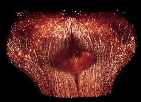
The Max Planck Institute of Neurobiology uses Amira software for axon tracing from head to toe
Ali Ertürk and his collaborators described a novel histo-chemical technique to clear spinal cord tissue of adult mice for fluorescence microscopy imaging of the intact spinal cord.
Previously, the high lipid content of the spinal cord tissue in adult mice allowed imaging of the spinal cord only through destructive histological methods, making the tracing of complete axons impossible. As applied in this study, the improved histo-chemistry enabled Ertürk and his colleagues to show that... Read more
Ali Ertürk, Christoph P Mauch, Farida Hellal, Friedrich Förstner, Tara Keck, Klaus Becker, Nina Jährling, Heinz Steffens, Melanie Richter, Mark Hübener, Edgar Kramer, Frank Kirchhoff, Hans Ulrich Dodt & Frank Bradke